
Cartilage Damage in Sydney
Understand the symptoms, causes, diagnosis and treatment options for knee cartilage damage, including non-surgical care and when cartilage repair may be considered.
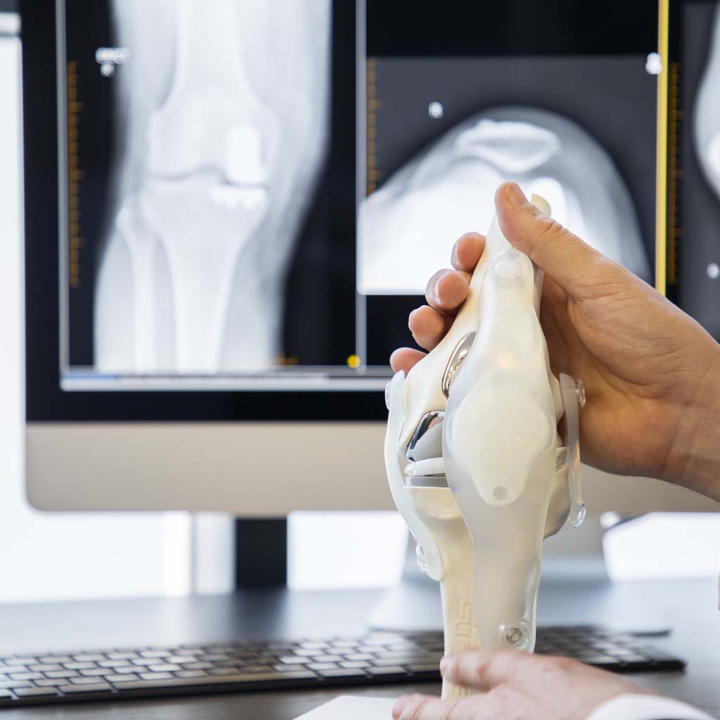
What is cartilage damage?
Cartilage damage is a common cause of knee pain, swelling and reduced joint function. It can affect people of all ages, from younger athletes who sustain a sporting injury to older adults experiencing gradual wear within the joint. Depending on the size and location of the injury, damaged knee cartilage may cause persistent discomfort, mechanical symptoms such as catching or locking, and difficulty with everyday activities.
Articular cartilage has a limited ability to heal naturally because it has very little direct blood supply. While many people improve with conservative care, some larger or persistent cartilage injuries may require specialist assessment to determine whether cartilage repair surgery or another treatment option may be appropriate. Early assessment can help guide management based on symptoms, examination findings and imaging results.
This page explains what cartilage damage is, its common causes and symptoms, how it is diagnosed, the available non-surgical and surgical treatment options and what recovery may involve. It also outlines when specialist assessment may be recommended.
If you are looking for information about other conditions affecting the hip or knee, visit the orthopaedic conditions page for further information.
Cartilage damage refers to injury or deterioration of the smooth protective cartilage covering the ends of the bones within a joint. In the knee, this specialised tissue is known as articular cartilage. It allows the femur, tibia and patella to glide smoothly over one another during movement while helping distribute forces across the joint.
Unlike muscle or skin, articular cartilage contains very few cells and has almost no direct blood supply. As a result, damaged cartilage has a limited ability to repair itself naturally. The extent of recovery often depends on the depth and size of the injury, as well as factors such as age, overall joint health and activity level.

Cartilage damage may range from small surface irregularities through to full-thickness cartilage defects where the protective layer has been completely lost. In some cases, the underlying bone may also become involved.
Symptoms do not always correspond directly with the size of a cartilage defect. Some people experience significant pain from relatively small injuries, while others with larger defects have only mild symptoms.
It is also important to distinguish cartilage damage from knee osteoarthritis. While both involve changes to the joint surface, isolated cartilage injuries often affect a specific area of otherwise healthy cartilage, whereas osteoarthritis usually involves more widespread changes throughout the joint.
Symptoms of cartilage damage
The symptoms of knee cartilage damage vary according to the location, size and depth of the injury. Associated problems involving the meniscus, ligaments, bone or kneecap can also influence the presentation.
Knee pain
Pain may develop during walking, running, squatting, climbing stairs or sporting activity. The location of discomfort can depend on which part of the joint surface is affected.
Swelling
Cartilage damage may irritate the knee joint and cause intermittent swelling. Swelling may increase after activity or following repeated loading through the affected area.
Catching or locking
Unstable cartilage or a loose fragment within the joint may cause catching, locking or a feeling that movement is being temporarily blocked. These symptoms may also occur with meniscal injuries and require appropriate assessment.
Clicking or grinding
Some people notice clicking, grinding or a rough sensation during knee movement. These symptoms do not necessarily indicate severe damage and should be considered alongside pain, swelling and function.
Reduced movement
Pain, swelling or mechanical irritation can make it difficult to fully bend or straighten the knee. Reduced movement may affect walking, sitting, kneeling, exercise and everyday activities.
Difficulty with work or sport
Cartilage injuries may reduce tolerance for running, jumping, pivoting, prolonged standing or physically demanding work. Symptoms may become more noticeable as the level or intensity of activity increases.
Seek prompt medical assessment. Sudden severe pain, inability to bear weight, a knee that remains locked, rapid swelling after injury or a hot and red joint should be assessed promptly.
What causes cartilage damage?
Cartilage damage may occur suddenly following an injury or develop gradually over time. In some cases, several factors contribute to deterioration of the joint surface.
Sporting injuries
Twisting, pivoting, landing or direct impact injuries can damage the articular cartilage. Cartilage injuries may occur alongside an anterior cruciate ligament tear, meniscal injury, kneecap dislocation or other knee trauma.
Direct impact
A fall or forceful blow to the knee can compress the joint surfaces and injure the cartilage or underlying bone.
Patellar instability
A dislocated or unstable kneecap can damage cartilage on the back of the patella or within the trochlear groove as the kneecap moves out of its normal position.
Repetitive loading
Repeated high-impact loading may contribute to symptoms when the joint surface has already been injured or is unable to tolerate the demands being placed upon it.
Knee alignment
Bow-legged or knock-kneed alignment can concentrate load through one part of the knee. This may affect the success of treatment and may need to be considered when planning cartilage preservation procedures.
Osteochondritis dissecans
Osteochondritis dissecans is a condition involving a segment of bone and overlying cartilage. The affected area may remain stable or become loose within the joint.
Gradual joint degeneration
Cartilage may deteriorate as part of osteoarthritis. Widespread arthritic cartilage loss is different from a localised cartilage defect and may require a different treatment approach.
How is cartilage damage diagnosed?
Diagnosis usually involves a detailed medical history, physical examination and appropriate imaging. The aim is to identify where the cartilage injury is located, determine its likely significance and assess whether other knee structures are involved.
Your clinician may ask how the symptoms began, whether there was a specific injury and which activities cause pain, swelling, catching or locking. Previous knee injuries, surgery and rehabilitation may also be reviewed.

Physical examination
The examination may assess swelling, tenderness, knee movement, alignment, stability, muscle strength and walking pattern. The clinician may also perform tests for meniscal, ligament or kneecap problems that could contribute to the symptoms.
X-rays
X-rays do not show articular cartilage directly, but they can identify joint-space narrowing, bone changes, alignment problems, previous fractures and signs of osteoarthritis.
MRI
MRI can provide detailed information about the cartilage, underlying bone, menisci and ligaments. It may help identify the size, depth and location of a cartilage defect and whether swelling or injury is present in the bone beneath it.
MRI findings must be interpreted alongside symptoms and physical examination. Not every cartilage abnormality seen on a scan is necessarily responsible for the patient's pain.
Arthroscopy
Arthroscopy allows the joint surfaces to be viewed directly using a small camera. It is not routinely required only to diagnose cartilage damage but may confirm the extent of an injury when arthroscopic treatment is being performed.
Non-surgical treatment options
For many people, surgery is not the first or only option for treating cartilage damage. The most appropriate approach depends on symptoms, the size and location of the cartilage defect, activity level and whether other knee structures have also been injured.
Surgery is not required for every cartilage injury. Many patients begin with rehabilitation, activity modification and symptom management. Treatment should be based on pain, function, examination findings, imaging and individual goals rather than the scan alone.
Activity modification
Activities that repeatedly aggravate the knee may need to be temporarily modified. This may include reducing running, jumping, deep squatting or other high-impact movements while maintaining suitable lower-impact activity.
Physiotherapy and strengthening
Physiotherapy for knee conditions may help improve muscle strength, balance, knee control and movement patterns while reducing unnecessary stress on the damaged area.
A rehabilitation programme may include quadriceps and hip strengthening, mobility work, balance exercises and a gradual return to work, recreation or sport. Exercises should be progressed according to symptoms and function.
Mobility and flexibility
Restoring or maintaining knee movement may help reduce stiffness and improve function. Exercises may also address restrictions involving the hip, ankle or surrounding muscles where relevant.
Pain-relieving medication
Pain-relieving or anti-inflammatory medication may be considered where medically appropriate. Suitability depends on medical history, other medicines and possible side effects and should be discussed with a GP, pharmacist or treating clinician.
Weight management
Where relevant, gradual weight management may reduce loading through the knee. Advice should be individualised and may involve support from a GP, dietitian or another qualified health professional.
Injection treatments
Injection treatment may occasionally be considered for symptom management in selected patients. The potential benefit varies, and injections do not restore normal articular cartilage. Their role, limitations and possible risks should be discussed with the treating clinician.
When might surgery be considered?
Cartilage repair surgery may be considered when a cartilage defect continues to cause significant pain, swelling or mechanical symptoms despite appropriate non-surgical management.
A surgical opinion may also be considered when:
- the cartilage defect is localised and suitable for repair or restoration;
- symptoms significantly affect work, exercise, sport or daily activities;
- the knee repeatedly swells following activity;
- catching or locking symptoms remain persistent;
- a loose cartilage or bone fragment is present;
- an associated ligament, meniscal or kneecap injury requires treatment;
- non-surgical treatment has not provided sufficient improvement; or
- the patient wishes to understand whether a cartilage preservation procedure may be appropriate.
The presence of cartilage damage on MRI does not automatically mean that surgery is required. Treatment decisions should consider the overall condition of the knee, symptoms, function, alignment, activity goals and the likelihood that a procedure may provide meaningful benefit.
How is cartilage repair surgery performed?
When non-surgical management has not provided sufficient improvement, cartilage repair surgery may be considered for carefully selected patients. The aim is to restore or improve the damaged joint surface, reduce symptoms and help preserve knee function where appropriate.
The specific procedure recommended depends on several factors, including the size and depth of the cartilage defect, its location within the knee, the quality of the surrounding cartilage and bone, age, activity level and associated injuries.
Before surgery
Before recommending surgery, the surgeon will review the patient's medical history, complete a physical examination and assess imaging such as MRI and X-rays. Additional investigations may occasionally be required to understand the condition of the knee fully.
The expected benefits, limitations, rehabilitation requirements and potential risks are discussed before a decision is made.
During surgery
Many cartilage procedures are performed arthroscopically using small incisions and specialised instruments. Depending on the injury, treatment may involve smoothing unstable cartilage, stimulating new repair tissue, transplanting healthy cartilage and bone or using specialised cartilage restoration techniques.
The surgical technique is selected according to the individual characteristics of the injury rather than applying a single approach to every patient.
Chondroplasty
Chondroplasty involves trimming or smoothing unstable cartilage. It may reduce mechanical irritation but does not restore the cartilage to its original condition.
Microfracture or marrow stimulation
Small openings are created in the bone beneath the defect to encourage blood and bone-marrow cells to form repair tissue. The new tissue differs from normal articular cartilage and may not be appropriate for every defect.
Osteochondral grafting
Healthy cartilage and underlying bone may be transferred from another area of the knee or provided through donor tissue. This may be considered for selected defects involving both cartilage and the underlying bone.
Cell-based cartilage restoration
Some cartilage injuries may be treated using specialised techniques involving cartilage cells and a supporting membrane or scaffold. Suitability depends on the defect, surrounding joint health and other patient factors.
After surgery
Following surgery, a structured rehabilitation programme is essential. Early movement is often encouraged while the repaired cartilage is protected. Depending on the procedure performed, temporary restrictions on weight-bearing or certain activities may be necessary.
Benefits and goals of treatment
The primary goal of cartilage damage treatment is to improve knee function while helping reduce pain and mechanical symptoms. Whether treatment is surgical or non-surgical, the objectives may include:
- reducing pain during daily activities;
- improving knee movement and flexibility;
- decreasing swelling and joint irritation;
- reducing catching or locking symptoms;
- supporting participation in work, recreation and sport where appropriate;
- helping preserve healthy joint surfaces where possible; and
- improving overall quality of life.
Every patient responds differently to treatment. Outcomes depend on factors such as the size of the cartilage defect, associated injuries, rehabilitation participation, age and the overall condition of the knee joint.
Risks and considerations
Non-surgical care is generally low risk, although symptoms may continue and activity may need to be modified while strength and function improve.
All surgical procedures carry potential risks. Risks and considerations associated with cartilage surgery may include:
- persistent pain or stiffness;
- incomplete healing of the repaired cartilage;
- infection;
- bleeding or haematoma;
- blood clots;
- ongoing swelling;
- failure to achieve the desired improvement;
- damage to surrounding structures;
- the need for additional surgery; and
- progression of joint degeneration over time.
Following the rehabilitation programme carefully and attending scheduled follow-up appointments are important parts of recovery, although a particular outcome cannot be guaranteed.
Recovery and rehabilitation
Recovery following cartilage damage treatment varies considerably depending on whether management is non-surgical or surgical, the severity of the injury and the type of procedure performed.
Recovery without surgery
Improvement with rehabilitation is usually gradual. Progress may be assessed through changes in pain, swelling, strength, walking tolerance and the ability to complete work, exercise or everyday activities.
Occasional symptom fluctuations can occur as activity levels change. The rehabilitation programme may need to be adjusted if pain or swelling increases substantially or persists.
Early recovery after surgery
During the early stages after surgery, treatment focuses on controlling swelling, protecting the healing tissues and gradually restoring knee movement. Some patients may require crutches or temporary restrictions on weight-bearing.
The length of the protected phase depends on the procedure performed, the size and location of the defect and the surgeon's rehabilitation protocol.
Physiotherapy and rehabilitation
Rehabilitation plays a central role in recovery. Progressive strengthening, balance training and movement retraining help restore knee function while reducing unnecessary stress on the healing joint surface.
The programme may include:
- swelling and pain management;
- gradual restoration of knee movement;
- quadriceps and hip strengthening;
- balance and neuromuscular training;
- walking and movement retraining;
- progressive loading of the knee; and
- preparation for work, recreation or sport.
Return to work
Returning to work depends on the physical demands of the occupation and the treatment performed. Office-based work may be resumed earlier than jobs involving prolonged standing, climbing, kneeling, lifting or heavy manual labour.
Driving
Driving should not resume until the patient can safely control the vehicle, enter and exit the car, complete an emergency stop and is no longer taking medication that affects alertness. Individual clearance should be obtained from the treating team.
Return to sport and activity
Returning to sport is based on recovery milestones rather than a fixed timeframe. Strength, balance, knee movement, swelling, confidence and the surgeon's assessment can all influence when higher-level activities may resume.
Returning too quickly may overload the healing joint surface. Progression should follow the recommendations of the surgeon and rehabilitation team.
How is cartilage damage different from other knee conditions?
Cartilage damage is only one possible cause of knee pain and reduced function. Ligament injuries, meniscal tears, tendon disorders and osteoarthritis may produce similar symptoms but often require different treatment approaches.
Cartilage damage and meniscal tears
Articular cartilage covers the ends of the bones, while the menisci are shock-absorbing structures positioned between the femur and tibia. Both may cause pain, swelling or catching, but they involve different tissues.
Cartilage damage and ligament injuries
Ligaments help stabilise the knee. A ligament injury may occur at the same time as cartilage damage following a twisting or sporting injury.
Cartilage damage and osteoarthritis
Unlike widespread knee osteoarthritis, cartilage damage frequently involves a localised defect within an otherwise relatively healthy joint. Osteoarthritis involves broader changes that may affect cartilage, bone, the joint lining, menisci and surrounding muscles.
Many people improve with rehabilitation alone, while selected patients with larger or persistent cartilage defects may benefit from cartilage preservation procedures. Treatment should reflect the individual diagnosis rather than imaging findings alone.
Questions to ask your surgeon
- What type of cartilage damage do I have?
- Where is the cartilage defect located?
- How large and deep is the damaged area?
- Are other knee structures also injured?
- Do I need surgery or could non-surgical treatment be appropriate?
- Would additional imaging be helpful?
- Which cartilage repair procedure, if any, may suit my injury?
- What are the expected benefits and limitations of treatment?
- What are the risks relevant to my circumstances?
- How long is rehabilitation likely to take?
- Will I need crutches or weight-bearing restrictions?
- When may I return to work, driving and sport?
- What can I do to help protect my knee in the future?
Assessment for cartilage damage in Sydney
Cartilage damage can affect knee comfort, movement and participation in work, exercise and sport. While some injuries improve with carefully supervised rehabilitation, others may require further investigation or consideration of cartilage repair procedures.
An accurate diagnosis can help determine whether symptoms are related to a cartilage defect, meniscal injury, ligament problem, kneecap condition, osteoarthritis or another source of knee pain.
Assessment may be appropriate when knee pain, swelling, catching or locking continues despite suitable care, when symptoms follow a significant injury or when the knee is limiting work, exercise or everyday activity.
Book an assessment with MTP Health in Sydney to discuss your knee symptoms, imaging and available non-surgical or surgical treatment options.
Your surgeon

Dr Jonathan Negus
Dr Jonathan Negus assesses and treats knee conditions, including articular cartilage injuries. Assessment considers symptoms, examination findings, imaging, knee alignment, associated injuries, previous treatment, general health and individual activity goals before any treatment recommendation is made.
View full profile →Frequently asked questions
Can cartilage damage heal naturally?
Small cartilage injuries may become less symptomatic with appropriate rehabilitation, but articular cartilage has a limited capacity to repair itself completely because it has very little direct blood supply. Treatment recommendations depend on the severity of the injury, associated knee problems and the patient's symptoms.
Is surgery always required for cartilage damage?
No. Many people improve with physiotherapy, strengthening exercises, activity modification and other conservative treatments. Surgery is generally considered when symptoms persist despite appropriate non-surgical care or when a specific cartilage defect may benefit from intervention.
How is cartilage damage diagnosed?
Diagnosis usually involves a detailed medical history, physical examination and imaging. MRI is commonly used to assess cartilage injuries, while X-rays can help evaluate bone alignment, previous injury and signs of arthritis.
Can I continue exercising with cartilage damage?
In many cases, yes. Low-impact exercise and a supervised rehabilitation programme may help improve strength and knee function. The most appropriate activities depend on the location and severity of the injury, symptoms and any associated knee conditions.
Will cartilage damage lead to arthritis?
Not necessarily. Some cartilage injuries remain stable for many years, while others may increase the risk of joint degeneration over time. The risk can depend on the size and location of the injury, knee alignment, associated damage and other individual factors.
How long does recovery take after cartilage repair surgery?
Recovery varies according to the procedure performed, the size and location of the cartilage defect, overall health and rehabilitation progress. Some procedures require a period of restricted weight-bearing followed by several months of progressive strengthening and activity-based rehabilitation.
Cartilage damage assessments across Sydney
North Shore Health Hub, Level 4, Suite 401, 7 Westbourne St, St Leonards NSW 2065
St Leonards consulting →173 Warringah Road, Beacon Hill NSW 2100 — serving the Northern Beaches
Beacon Hill consulting →Also consulting at Gosford, Wahroonga, Castle Towers and Tamworth.
Discuss your knee symptoms with MTP Health
Book an assessment to clarify the cause of your symptoms and discuss appropriate non-surgical and surgical treatment options.
Book an AssessmentPrefer to talk? Call (02) 9437 9794 · GP & physio referrals: referrer information