
Distal Femoral Osteotomy in Sydney
A distal femoral osteotomy corrects alignment at the thigh bone, most often for knock-knee patterns that overload the outer knee. By shifting load toward a healthier part of the joint, it can reduce pain, protect cartilage and help delay the need for knee replacement in carefully selected patients.
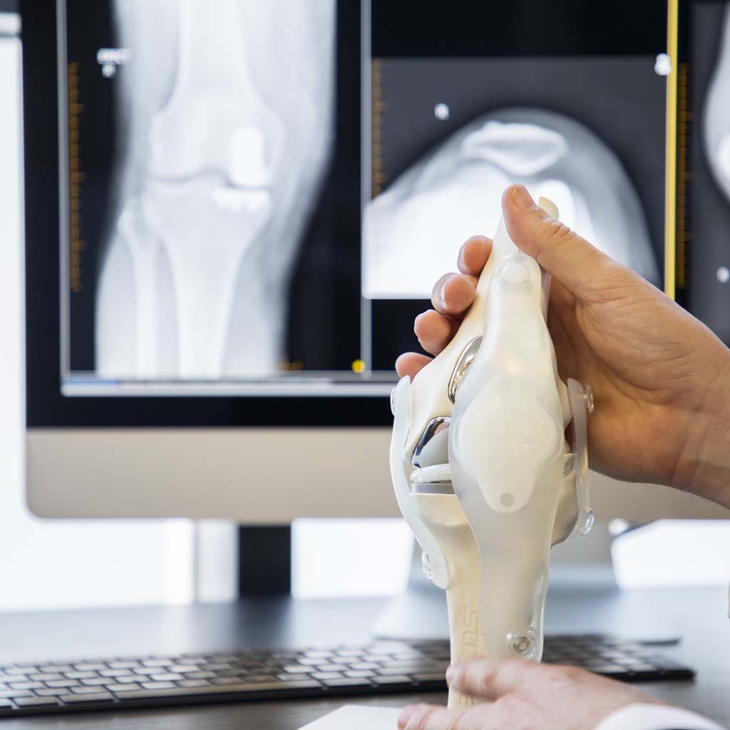
What is a distal femoral osteotomy?
A distal femoral osteotomy, often shortened to DFO, is a knee realignment operation performed at the lower end of the thigh bone, just above the knee. It is most often used when valgus alignment, commonly called knock-knees, places too much load through the outer, or lateral, side of the knee.
The aim is not to replace the knee joint. Instead, the bone is carefully cut, realigned and fixed in a new position so that weight passes through the knee in a more balanced way. This can reduce pressure on the overloaded lateral compartment and help protect the joint when the rest of the knee is still suitable to preserve.
A distal femoral osteotomy is a more selective procedure than a knee replacement. It is a type of knee osteotomy usually considered for younger or more active patients, or for people whose knee wear is mainly on one side of the joint. A high tibial osteotomy may be considered when realignment is needed through the tibia instead. It may also be used alongside cartilage restoration, meniscal transplant or ligament surgery when alignment is contributing to the problem.

Do you need a distal femoral osteotomy?
A distal femoral osteotomy is usually considered when valgus alignment is contributing to overload on the outside of the knee. This may cause lateral knee pain, swelling after activity, reduced walking tolerance, difficulty on uneven ground or symptoms that keep returning despite good rehabilitation.
You may be suitable if your symptoms match your imaging, the lateral compartment is overloaded, and the rest of the knee has enough healthy cartilage to preserve. Assessment usually includes a detailed history, knee examination, standing long-leg X-rays and often MRI to understand the cartilage, meniscus and ligament picture.
Alignment matters, but symptoms matter too. Not every knock-knee needs surgery. Some people have valgus alignment without pain or joint damage. Others improve with strength work, activity modification, bracing or physiotherapy. Our physiotherapy team, exercise physiologists and orthopaedic surgeons work together to help you understand whether your alignment is truly driving your symptoms.
A distal femoral osteotomy may not be suitable if knee osteoarthritis is widespread across the knee, the joint is very stiff, inflammatory arthritis is present, the knee is severely unstable, bone quality is poor, or a knee replacement would provide a more reliable result. The goal of your consultation is to match the treatment to the actual problem, not simply to the appearance of the leg.
Benefits and risks
What you can expect it to achieve
- Reduce load through the overloaded lateral compartment of the knee
- Improve pain related to valgus alignment and outer-knee overload
- Help preserve the natural knee joint in suitable patients
- Potentially delay the need for total knee replacement
- Support other joint-preserving procedures, such as cartilage or meniscus surgery, when alignment is part of the problem
- Improve lower-limb mechanics for walking, stairs and uneven ground
- Allow a personalised rehabilitation plan focused on strength, control and confidence
Risks to understand
- Common and temporary: swelling, bruising, stiffness, discomfort around the plate and reduced strength in the early recovery period
- Bone healing risks: delayed union, non-union, loss of correction or fracture through the bone hinge
- Surgical risks: infection, wound problems, blood clots, nerve or blood vessel injury, and anaesthetic complications
- Hardware-related risks: plate or screw irritation, screw problems or the need for later removal if hardware becomes symptomatic
- Longer-term risks: ongoing pain, progression of arthritis, incomplete symptom relief or later conversion to knee replacement
All surgery carries risk and outcomes vary between individuals. A distal femoral osteotomy can be very useful when alignment is the main driver of lateral knee overload, but it is not the right operation for every painful knee.
Distal femoral osteotomy vs knee replacement
The main difference is that a distal femoral osteotomy preserves the knee joint, while a knee replacement resurfaces or replaces arthritic joint surfaces. A DFO changes the way load passes through the knee. A replacement changes the worn joint surfaces themselves.
For younger, active patients with valgus alignment and mainly lateral compartment overload, realignment may help protect the joint and delay replacement. If arthritis is advanced across several parts of the knee, a total knee replacement may be more appropriate.

| Distal femoral osteotomy | Knee replacement |
|---|---|
| Realigns the thigh bone to redistribute load | Replaces worn joint surfaces with implants |
| Preserves the natural knee joint | Used when joint surfaces are too damaged to preserve |
| Usually considered for selected active patients | Often used for advanced arthritis affecting daily life |
| Requires bone healing before full progression | Usually allows earlier weight-bearing, depending on the case |
| May delay the need for replacement | Designed to treat established end-stage arthritis |
The procedure: what happens
Before surgery, your surgeon will plan the correction using standing long-leg X-rays. These images show the hip, knee and ankle together, allowing the mechanical axis of the leg to be measured. MRI may also be used to assess cartilage, meniscus and ligament health.
The operation is performed under general or spinal anaesthesia. During surgery, the distal femur is exposed through an incision around the lower thigh and knee. The bone is carefully cut in a controlled way and then opened or closed depending on the correction required.
There are two main techniques: a lateral opening wedge osteotomy and a medial closing wedge osteotomy. In an opening wedge procedure, a controlled gap is created and may be supported with bone graft or bone substitute. In a closing wedge procedure, a wedge of bone is removed and the bone ends are brought together. The best technique depends on your anatomy, correction size, bone quality and your surgeon’s plan.
Once the alignment has been corrected, the bone is fixed with a plate and screws. X-ray imaging is used during the operation to check the correction and hardware position. The wound is then closed and covered with dressings.
After surgery
You will wake in the recovery room, where your pain, circulation and vital signs are monitored closely. Once stable, you will return to the ward and begin early recovery with nursing and physiotherapy support.
Unlike many knee replacement procedures, full weight-bearing is not always immediate after a distal femoral osteotomy. Because the bone needs to heal, weight-bearing is usually progressed gradually and guided by your surgeon, X-rays and rehabilitation team.
When to seek help. Contact us or seek urgent care if you develop a fever, increasing calf pain or swelling, spreading redness, wound discharge, shortness of breath, chest pain, sudden worsening pain, new numbness or pain that is not controlled by medication. Concerned about your recovery? Call (02) 9437 9794.
Distal femoral osteotomy recovery and rehabilitation
Recovery after distal femoral osteotomy is built around two priorities: allowing the bone to heal and restoring strength, movement and confidence. Because the procedure creates a controlled bone cut, your recovery is usually more protected than after many soft-tissue knee operations.
You will usually use crutches after surgery. Weight-bearing is often limited at first, then increased gradually over several weeks once follow-up X-rays show appropriate healing. The exact plan depends on the technique used, the amount of correction, bone quality, fixation strength and your surgeon’s instructions.
Recovery timeline
| Phase | Timeframe | What to expect |
|---|---|---|
| Protect & settle | Days 0–14 | Crutches, swelling control, wound care, gentle movement, pain management and early muscle activation |
| Protected loading | Weeks 2–6 | Gradual rehabilitation while protecting the osteotomy site; weight-bearing depends on your surgeon’s protocol |
| Healing check | Weeks 6–12 | X-rays guide progression; walking, range of motion, strength and balance work are gradually increased |
| Strength & control | Months 3–6 | Progressive strengthening, gait retraining, cycling, balance and return to more normal daily activity |
| Return to activity | Months 6–12 | Ongoing strength, confidence and conditioning; return to higher-level activity is individual and surgeon-guided |
Timeframes are a guide. Your surgeon and rehabilitation team will progress you based on bone healing, symptoms, strength and your goals.
Post-operative care
At home, the priorities are protecting the osteotomy, keeping the wound clean and dry, controlling swelling, using crutches safely, taking medication as prescribed and completing your rehabilitation exercises. Follow-up X-rays are used to monitor bone healing before your loading and activity levels are increased.
Rehabilitation at MTP Health
This is where MTP Health is different. Your rehabilitation is delivered by our own physiotherapists and exercise physiologists, working in the same clinic as your surgeon. Your plan can begin before surgery and continue afterwards, with everyone working from the same goals and shared clinical record.
Rehabilitation focuses on restoring knee movement, rebuilding quadriceps and hip strength, improving balance, retraining walking mechanics and preparing you for the activities that matter to you. Your recovery may be supported by our pre-operative rehabilitation, post-operative rehabilitation, physiotherapy and exercise physiology services.
Returning to driving, work and sport
Return to driving depends on which leg was operated on, whether you drive an automatic or manual car, your weight-bearing status, reaction time, pain control and medication use. You must be able to safely perform an emergency stop and must no longer be taking strong pain medication. Please check with your surgeon and insurer before driving.
Desk-based work may be possible once pain is controlled, mobility is safe and you can manage transport and swelling. More physical work usually takes longer, especially if it involves standing, lifting, stairs, kneeling or uneven ground. Return to sport is gradual and depends on bone healing, strength, joint health and the type of activity.
Rehab is part of the operation. A distal femoral osteotomy changes your alignment, but the result depends heavily on bone healing, strength and movement quality. The best outcomes come from combining careful surgical correction with a structured, staged rehabilitation plan.
Distal femoral osteotomy cost in Sydney
The cost of distal femoral osteotomy depends on your private health cover, hospital insurance, surgeon fees, anaesthetist fees, assistant fees, hospital charges, implants, imaging and any excess on your policy.
If you are having surgery using private health cover, your out-of-pocket costs will depend on your level of cover, your fund’s arrangements, the hospital used and whether any reduced-gap arrangements apply. The anaesthetist is an independent practitioner and may charge a separate gap, so we provide their details and recommend checking their quote before surgery.
Because DFO surgery can involve specialised planning, implants and a longer rehabilitation pathway than some knee procedures, your team will talk you through expected costs before surgery wherever possible.
If surgery is performed through the public system, there may be no out-of-pocket surgical cost, but waiting times can be long and depend on hospital access, urgency category and local availability.
Why have your distal femoral osteotomy at MTP Health
MTP Health brings together fellowship-trained orthopaedic surgeons, careful surgical planning, physiotherapy and exercise physiology in one clinic. That matters because a distal femoral osteotomy is not just about changing an X-ray angle — it is about selecting the right patient, planning the correction accurately and guiding recovery properly.
Honest advice comes first. If your knee is better suited to non-surgical care, we will tell you. If arthritis is too advanced for realignment, we will explain why a replacement option may be more reliable. If a distal femoral osteotomy is suitable, we will help you understand the procedure, recovery, risks and expected outcome clearly before you decide.
Your surgeon

Dr Jonathan Negus
Dr Negus is a fellowship-trained hip and knee surgeon with a particular focus on joint preservation and joint replacement decision-making. He works alongside MTP Health's physiotherapy and exercise physiology team so your surgery and rehabilitation are managed as one plan.
View full profile →Frequently asked questions
What symptoms can a distal femoral osteotomy help with?
A distal femoral osteotomy is usually considered when valgus alignment is loading the outer, or lateral, side of the knee. Common symptoms include discomfort when weight-bearing, reduced walking tolerance, swelling after activity, trouble on uneven ground or pain that keeps returning despite good non-surgical care.
Whether it suits you depends on your imaging findings, the location of your pain, your cartilage health, ligament stability and the overall condition of the joint.
How long does the bone take to heal?
Bone healing varies from person to person. Weight-bearing is often progressed gradually over several weeks, with X-rays used to check healing at follow-up appointments.
Strengthening usually continues for several months. Bone quality, smoking status, the amount of correction, fixation stability and how rehabilitation progresses all play a part.
Can a distal femoral osteotomy delay the need for a knee replacement?
Yes, in suitable patients it may help delay the need for knee replacement. By shifting load toward the healthier part of the knee, the procedure can help preserve the joint when wear affects mainly one compartment.
How long that benefit lasts differs between people and depends on cartilage health, alignment, activity levels, body weight, rehabilitation and future wear patterns.
Will the plate and screws need to be removed?
The plate and screws usually stay in place long term. Removal is not routine and is generally only considered if the hardware becomes uncomfortable, irritates nearby soft tissue or interferes with later treatment.
If hardware removal is considered, your surgeon will usually want to confirm that the osteotomy has fully healed before making that decision.
What is the difference between valgus alignment and knock-knees?
Valgus alignment describes an inward angle of the knee relative to the hip and ankle. “Knock-knees” is the everyday term for the same appearance.
Not every valgus knee causes problems, but a more pronounced inward angle can place extra load on the outer side of the knee. Assessment helps clarify whether your alignment is contributing to your symptoms.
Is distal femoral osteotomy the same as knee replacement?
No. A distal femoral osteotomy is a joint-preserving realignment procedure. It changes the angle of the thigh bone to redistribute load through the knee.
A knee replacement resurfaces or replaces damaged joint surfaces. The right option depends on your age, activity goals, alignment, arthritis pattern and the overall health of the knee.
What imaging is needed before distal femoral osteotomy?
Standing long-leg X-rays are usually needed because they show the hip, knee and ankle together and allow your surgeon to measure the mechanical axis of the leg.
Standard knee X-rays and MRI may also be used to assess cartilage, meniscus, ligament health and whether arthritis is isolated enough for a joint-preserving procedure.
What are the main risks of distal femoral osteotomy?
The main risks include infection, blood clots, wound problems, nerve or blood vessel injury, stiffness, delayed bone healing, non-union, loss of correction, hardware irritation and ongoing pain.
Some people may still develop progressive arthritis over time and later need a knee replacement. Your surgeon will explain your individual risk profile before surgery.
Distal femoral osteotomy consultations across Sydney
North Shore Health Hub, Level 4, Suite 401, 7 Westbourne St, St Leonards NSW 2065
St Leonards consulting →173 Warringah Road, Beacon Hill NSW 2100 — serving the Northern Beaches
Beacon Hill consulting →Also consulting at Gosford, Wahroonga, Castle Towers and Tamworth.
Talk to a surgeon about your knee alignment
Book a consultation to find out whether a distal femoral osteotomy is right for you — and if it is not, to leave with a clear plan for the most suitable next step.
Book a ConsultationPrefer to talk? Call (02) 9437 9794 · GP & physio referrals: referrer information
- Current peer-reviewed literature on distal femoral osteotomy indications, techniques, survivorship and complications.
- Recent systematic reviews and cohort studies on lateral opening wedge and medial closing wedge distal femoral osteotomy for valgus alignment and lateral compartment overload.